normal range echocardiography normal values pdf
132 - 148 148. Right apical short axis with cursor at level of chordae attachment to papillary muscles.

Normal Values Of Echocardiographic Measurements A Population Based Study
67 yo male with SOB.

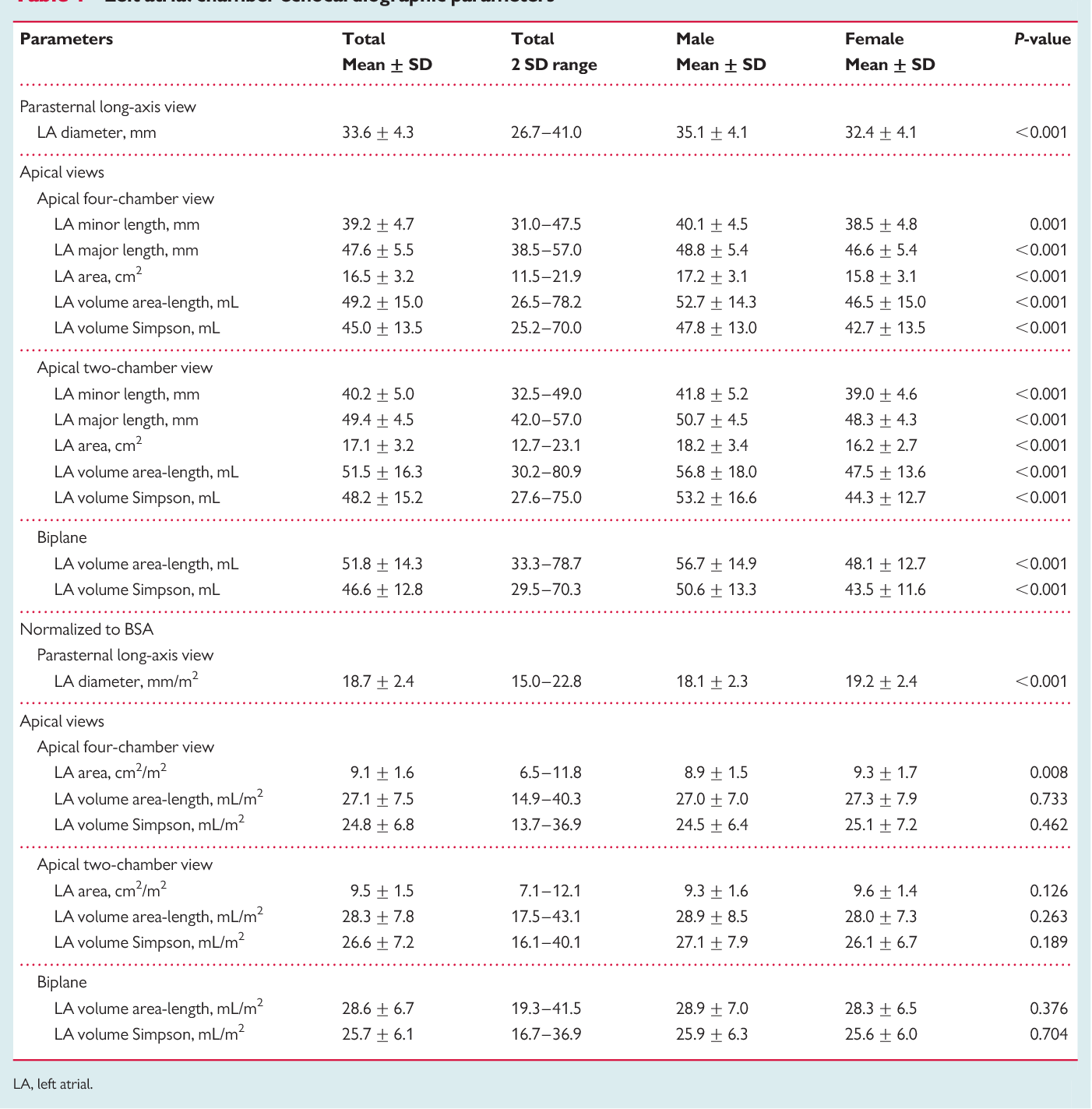
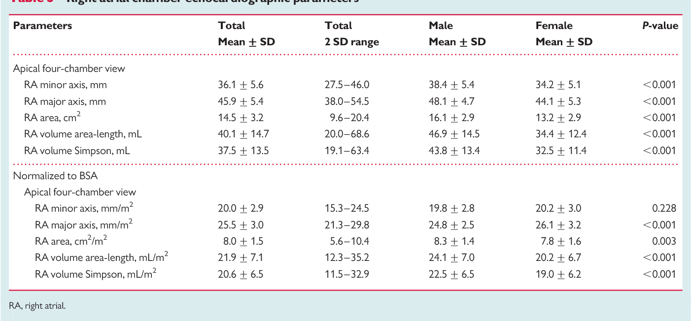
. Normal reference ranges for cardiac chambers size obtained in a large group of healthy volunteers accounting for gender and age highlight the need for body size normalization that should be performed together with age-and gender-specific assessment for the most echocardiographic parameters. Method to measure the distance of systolic excursion of the RV annular segment along its longitudinal plane from a standard apical 4- chamber window. 5 Mitral valve M-mode a.
Reference limits for echocardiography 7 1 G4 However for almost a century 196 has been rounded to 2 which covers 954 of the population 12. Conclusion The NORRE study provides normal values of proximal aorta dimensions as assessed by echocardiography. A normal ejection fraction is 53-73 52-72 for men 54-74 for women.
Figure A5M-mode end-diastolic ED septal thickness ST. Manual of Echocardiography. Normal EF was 63 6 5 using the biplane method of disks.
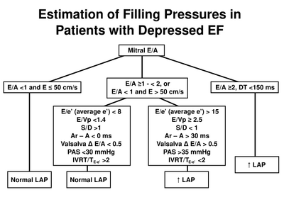
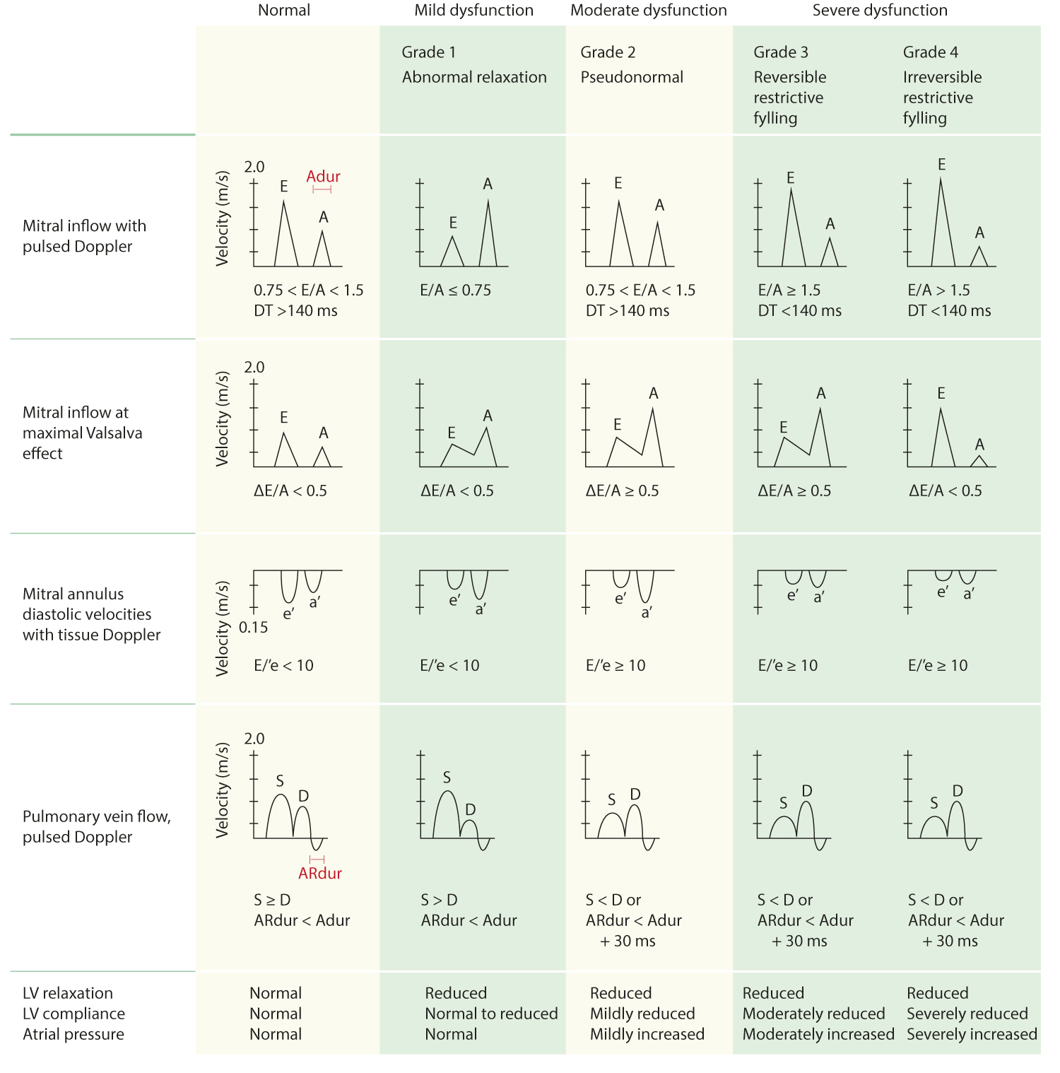
Refer to Table 2 normal values for non-contrast images and Table 4 recommendations for the normal range mildly moderately and severely abnormal ejection. Normal EA 10-20 2. The aim of the Normal Reference Ranges for Echocardiography Study NORRE Study is to obtain a set of normal values for cardiac chamber geometry and function in a large cohort of healthy Caucasian individuals aged over a wide range of ages 25-75 years using both conventional and advanced echocardiographic techniques.
The base of the RV free wall provides one of the most visibly obvious movements on normal echocardiography. LVIDD men 4259 cm LVIDD women 3953 cm LVEDV 46106 mL women LVEDV 62150 mL men LVESV 1442 mL women LVESV 2161 mL men RV FAC 35 LVOT stroke distance for intravascular volume status Ristow et. Reference ranges for different anatomical levels using different i measurement conventions and ii at different times of the cardiac cycle ie.
LV Dimensions Volumes Mass Normal Mild Moderate Severe Normal Mild Moderate Severe LVIDdiastolemm 3756 5761 6265 65 3551 5255 5659 59 LVIDsystolemm 2241 4245 4650 50 2037 3842 4346 46. Mid-systole and end-diastole are provided. Upper reference values mean 2 SD for the LV mass were 1041 gm 2 in men and 1001 gm 2 in women for ejection fraction were 713 in men and 726 in women for LV end-diastolic volume 757 and 676 mLm 2 for LV end-systolic volume 288 and 259 mLm 2 and for LV end-systolic dimension 207 and 213 mmm 2 respectively.
1 Left Ventricle M-mode and 2D a. Table 2 presents the absolute and indexed normal reference ranges of echocardiographic chamber dimensions in young eastern Indian adults as per this study. However due to the lack of consistency in current echocardiographic reference values their use for clinical decision-making remains questionableAimsThe aim of the Normal Reference Ranges for.
Normal range echocardiography normal values pdf. A TAPSE cutoff value 17 mm yielded high specificity though low sensitivity to. Diameter or A normal fetal heart rate FHR usually.
Pdf Echocardiographic Reference Ranges For Normal Cardiac Chamber Size Results From The Norre Study Semantic Scholar 458 133 years healthy volunteers 320 men and 414 women were enrolled at 22 collaborating institutions of the Normal Reference Ranges for Echocardiography NORRE. Upper normal reference limit UNRL OF LV end diastolic dimension LVEdD LVEDV LVESV indexed LVEDV iLVEDV iLVESV were slightly more for men while normal reference. Therefore in individuals aged 20 years EF in the range of 53 to 73 should be classified as normal.
Access Free Normal Reference Ranges For Echocardiography Classification of left ventricular size. Normal Echocardiographic Values for Cardiovascular Structures 885 05 10 15 20 25 M-mode ES PWT cm 00 05 10 15 20 25 Body Surface Area m2 2SD 2SD Mean Figure A4M-mode left ventricular end-systolic ES posterior wall thickness PWT versus body surface area. Three-dimensional Table 2 Normal values for 2D echocardiographic parameters of LV size and function according to gender.
Normal Dog 20 30 circsec Normal Cat 27 43 circsec. AIMS Availability of normative reference values for cardiac. Normal Ranges for LV Size and Function Normal values for LV chamber dimensions linear volumes and ejection fraction vary by gender.
Right apical short or long axis.
Pdf Normal Echocardiographic Measurements In Indian Adults How Different Are We From The Western Populations A Pilot Study
Pdf Echocardiographic Reference Ranges For Normal Cardiac Chamber Size Results From The Norre Study Semantic Scholar
Normal Values Of Tte Echopedia
Reference Normal Values For Echocardiography Ecg Echo

Pdf Echocardiographic Reference Ranges For Normal Cardiac Chamber Size Results From The Norre Study Semantic Scholar

Pdf Echocardiographic Reference Ranges For Normal Cardiac Doppler Data Results From The Norre Study

Pdf Echocardiographic Reference Ranges For Normal Cardiac Doppler Data Results From The Norre Study
Pdf Echocardiographic Reference Ranges For Normal Cardiac Chamber Size Results From The Norre Study Semantic Scholar

Normal Values Of Echocardiographic Measurements A Population Based Study

Pdf Echocardiographic Reference Ranges For Normal Cardiac Doppler Data Results From The Norre Study

Pdf Echocardiographic Reference Ranges For Normal Cardiac Chamber Size Results From The Norre Study Semantic Scholar

Normal Reference Values Of Echoca Preview Related Info Mendeley

Pdf Echocardiographic Reference Ranges For Normal Cardiac Doppler Data Results From The Norre Study Semantic Scholar

Pdf Normal Reference Intervals For Cardiac Dimensions And Function For Use In Echocardiographic Practice A Guideline From The British Society Of Echocardiography Semantic Scholar

Normal Values Of Echocardiographic Measurements A Population Based Study

Pdf Echocardiographic Reference Ranges For Normal Cardiac Chamber Size Results From The Norre Study Semantic Scholar
Normal Values Of Tte Echopedia

Pdf Echocardiographic Reference Ranges For Normal Cardiac Doppler Data Results From The Norre Study
Tissue Doppler Derived E E Ratio As A Parameter For Assessing Diastolic Heart Failure And As A Predictor Of Mortality In Patients With Chronic Kidney Disease